Microstructured CsI for
high resolution X-ray imaging
Home / Technologies / Caesium Iodide
An Introduction to Caesium Iodide
Digital imaging is rapidly advancing – particularly in dental diagnosis – with one of the major imaging techniques being based on scintillator coated fibre optic face plates (FOPs) bonded to CCD or CMOS devices.

Key to successful diagnosis are conflicting technical requirements which demand scintillator optimisation.
These include:
- high resolution imaging for diagnosis with high efficiency to minimise dose
- high X-ray absorption for improved signal:noise but preferential absorption of low energy X-rays for soft tissue diagnosis
- low granularity and high homogeneity of imaging field
- slim, convenient packaging which is robust, safe and has long life
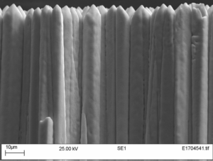
Scintacor has extensive experience in the custom development and production of optimised scintillator layers. Using Fiber Optic Plates (FOPs) which have high X- ray absorption, (thus protecting the CCD for extended life) Scintacor provides X-ray Fibre Optic Scintillators (FOS) for bonding and directly deposits phosphors and CsI:Tl for use in dental and other similar applications. Micro-columnar CsI:Tl grown on FOPs has been particularly successful at providing high resolution digital dental images. An SEM image of the CsI is shown in figure 2, showing the micro columnar structure of the coating that retains the high-resolution image – each column acting as an individual light-guide.

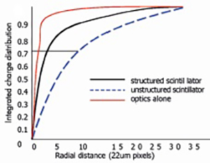
Comparing competitive bonded X-ray screens of Gadox:Tb phosphor and columnar CsI:Tl FOS shows the benefits conferred by columnar growth. The resolution performance demonstrated throughout the MTF curve in figure 1 shows CsI:Tl to have significant advantages with ultimate resolution being >18 line pairs/mm as opposed to the 12-1 4 lp/mm provided by the Gadox:Tb layer. Further, while Gadox:Tb has good efficiency, this is derived preferentially from the higher energy X-rays – CsI:Tl’s efficiency is derived at lower energies thus providing enhanced soft tissue imaging.

Other phosphors such as Y2O2S:Tb do absorb at low energies but are insufficiently dense to provide the high quality particulate layers and hence imaging performance seen from Scintacor’s columnar CsI:Tl depositions.
At Scintacor we continuously develop our product range and are able to deposit structured CsI onto a variety of substrates including glass, aluminium, and silicon sensors. By selecting the process used, we can provide single piece CsI scintillators up to 310mm x 310mm.
Scintacor has the knowledge and expertise based on years of experience to partner with you in the development of custom CsI:Tl products for dental, mammography and other X-ray imaging applications.
contact us for further information
Contact Us
+44 (0)1223 223 060
info@scintacor.com
